X ray machine working pdf
X ray exposure How safe are X rays Medical News Today December 18th, 2018 – X rays are a vital imaging tool used around the globe Since first being used to image bones over 100 years ago the X ray
X Ray Machine Working [Read Online] X Ray Machine Working Books Scan X Security Security X Ray Machines Metal Detection December 7th, 2018 – Scan X …
The x ray machine is a device for producing X light, which is mainly composed of the X light ball tube and the power supply of the X ray machine and the control circuit. When the power is turned on, the start button of the x ray machine is pressed and the whole machine starts to work.
X-ray machines work by generating an electrical current or voltage, which is then projected through an X-ray tube to produce a series of X-ray waves, which either pass through objects or are absorbed by the surrounding material.
beam of panoramic dental x-ray machines is fully intercepted by the image detector, it is not necessary to identify adjacent occupied areas in the workload information for this type of machine.
It takes a different form of X-ray and uses lower doses of radiation than a usual X-ray. Because these X-rays do not go through breast tissue as easily, the mammogram machine has two plates that compress the breast to spread the tissue apart. A more accurate image is obtained with less radiation this way.
RADIATION INFORMATION FOR HOSPITAL PERSONNEL 1. INTRODUCTION X-ray machines and radiation emitting sources are used in hospitals for the diagnosis and treatment of diseases.
How does the equipment work? The linear accelerator uses microwave technology (similar to that used for radar) to accelerate electrons in a part of the accelerator called the “wave guide,” then allows these electrons to collide with a heavy metal target to produce high-energy x-rays.
target to produce high-energy x-rays. These high energy x-rays are shaped as they exit the machine to These high energy x-rays are shaped as they exit the machine to conform to the shape of the patient’s tumor and the customized beam is directed to the patient’s tumor.
20/03/2014 · X-ray producing equipment and its location must be registered with Safety and Health and the Radiological Council, and x-ray machines may only be used by, or under the supervision of, a person with an appropriate licence.
24/08/2015 · An x-ray tube consists of an anode and a cathode within a casing that can hold vacuum. The cathode is heated to high temperatures, where it starts emitting electrons – this process is …
An X-ray machine is like a giant camera that allows doctors to see what is going on inside a patient without having to do surgery. To produce an X-ray picture, an X-ray machine produces a very concentrated beam of electrons known as X-ray photons.
Linear Accelerator RadiologyInfo.org
How does x-ray imaginig work? Xray Imaginis The
corresponding to the X-ray intensity distribution; electrons of this distribution are accelerated and focused on a secondary screen creating a reversed and reduced image with 100 to 1000 higher image intensity which can be observed by an
MEDICAL NOW No.70 (2011.8) RAD Digital Mobile X-Ray System with Wireless FPD MobileDaRt Evolution Medical Systems Division, Shimadzu Corporation
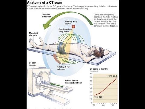
The term “computed tomography”, or CT, refers to a computerized x-ray imaging procedure in which a narrow beam of x-rays is aimed at a patient and quickly rotated around the body, producing signals that are processed by the machine’s computer to generate cross-sectional images—or “slices”—of the body.
2 During the past years we have increased our sales of radiometric Vollmer strip thickness measurement sys-tems, i.e. X-ray or isotope gauges, dramatically.
Working knowledge of an X-ray machine control panel (timer, relationship to generator and X-ray tube) Working knowledge of an X-ray tube (construction, components, operation) Essential skills: It is critical that the candidate demonstrate the ability to effectively do the task outlined in elements and performance criteria of this unit, manage the task and manage contingencies in the context of
X-rays are like radio waves and visible light electromagnetic radiation. X-rays, however, X-rays, however, have higher frequency, n , and shorter wavelength, l , than light and radio waves.
There are three main parts of an x-ray machine. The first is the x-ray tube. This is responsible for the creation of x-rays. Therefore, without this functioning properly, there would be little chance for the x-ray to work in the right way. It generates enough x-rays so that when they are sent toward
4 X-Ray Safety Manual 1.0 INTRODUCTION 1.1 Purpose of the Manual The Winnipeg Regional Health Authority (WRHA) X-Ray Safety Committee (XRSC) has undertaken the development of this manual to provide a common basis for
x ray machine working Fri, 07 Dec 2018 03:29:00 GMT x ray machine working pdf – An X-ray tube is a vacuum tube that converts electrical input power into X-rays.
ray inspectors and facilities with x-ray machines. No conclusions are included, and the No conclusions are included, and the implementation and use of the information contained in this document are solely the

the users are working near the X-ray equipment, the X-ray shutters are closed and no X-rays are emitted from the system. Only by closing the lead impregnated plastic doors and activating the
A PORTABLE CABINET X-RAY MACHINE TO CONTROL INSECTS IN EXPORTED FRUIT Peter Follett and Randy Kirk USDA-ARS, Hilo, Hawaii peter.follett@ars.usda.gov
Tender – Mobile X-ray Machine AIIMS-Jodhpur Page 2 All India Institute of Medical Sciences (AIIMS), Jodhpur, Rajasthan, an apex healthcare institute being
An X-ray tube is a vacuum tube that converts electrical input power into X-rays. X-ray tubes evolved from experimental Crookes tubes with which X-rays were first discovered on November 8, 1895, by the German physicist Wilhelm Conrad Röntgen.

Figure 2: Block Diagram of X-Ray Operation/Working of X-Ray Machine High voltage source and high voltage transformer . High voltage source is responsible for providing high voltage to the H.V transformer for a decided time. The H.V transformer produces 20 KV to 200 KV at the O/P. These voltages are used to determine the contrast of the image. High voltages have higher contrast. High voltage
An x-ray examination is used to create images of your internal organs or bones to help diagnose conditions or injuries. A special machine emits (puts out) a small amount of ionising radiation.
X-rays are also referred to as radiographs or roentgenograms (after W.C. Roentgen). Conventional x-ray imaging has evolved over the past 100 years, but the basic principal is still the same as in 1895.
X-ray equipment Safety Health and Wellbeing The
If x-rays travelling through the body also pass through an x-ray detector on the other side of the patient, an image will be formed that represents the “shadows” formed by the objects inside the body.
A guide to machinery and equipment safety is provided under the Work Health and Safety Act 2011 (x-rays, microwaves) Electrical Woodworking dust generated by a buzzer is removed via forced extraction and ventilation. Welding fumes are extracted via flexible, locatable forced extraction and ventilation system. PN10596 Version 2 Last updated August 2015 – Guide to machinery and equipment
Using X-ray vision to detect unseen gold 8 August 2013 An eight kilogram gold nugget. Powerful X-rays can now be used to rapidly and accurately detect gold in ore samples, thanks to a
X-rays work in exactly the same way. Bones and denser structures create a “shadow” on the X-ray film, and these shadows are used to by doctors to see breaks or other abnormalities in your bones. Bones and denser structures create a “shadow” on the X-ray film, and these shadows are used to by doctors to see breaks or other abnormalities in your bones.
FOR INTERNAL AFP USE ONLY AFP National Guideline on safe working with radiation 1. Disclosure and compliance This document is classified UNCLASSIFIED and is …
x ray machine ppt 1. X-RAY MACHINE 2. The extent of utilization (is the machine working), Severity of utilization (is the machine work under normal load condition), Operating condition, Are there any other specific factors that may affect the instrument 26.
X-rays are produced when high energy charged particles are rapidly decelerated or turned. X-ray production is the opposite of the photoelectric effect.
Typical X-ray machine 1950s Restriction of X-ray field Collimation X-Ray machine with slide in lead diaphragms. 2 Light (projection) beam diaphragm with adjustable Lead Shutters X-RAY MACHINES Portable max output 20mA, 80KV 50 mA 100KV High Frequency X-RAY MACHINES Ceiling mounted, 2000mA 150KV Automatic Film Processors 1960s Dry, Processed Film in 90 seconds Courtesy … – x wing templates dimensions pdf Medical Applications of X Rays by OTHA W. LINTON. 26 SUMMER 1995 A CENTURY OF RADIOLOGY: 1895–1995 The discovery of the X ray in 1895 was one of the most momentous events in science and medicine, but it was only the beginning of what was to be accomplished in the next 100 years in radiology. What follows are some highlights provided by American College of Radiology. …
X-ray machine and can reveal objects inside the body. 1 HOW MUCH RADIATION ARE PEOPLE EXPOSED TO IN X-RAY SCANNERS? When exposed to X-rays our body absorbs energy, the amount of energy effectively absorbed over time is expressed in “sievert” (Sv). Over the course of one year, a person should not be exposed to more than a total of 1 millisievert from man-made sources such as …
components of the x-ray machine. Today’s discussion will focus on the x-ray tube. In the picture, you can see the technologist handling the x-ray tube. She is manipulating the field size by adjusting a device called the “collimator” which is attached to the x- ray tube housing. The rectangular looking device just above the collimator is the housing for the x-ray tube. The diagram on the
• X-ray machine sends individual x-ray particles through the body. • The images are recorded on a computer or film. • The images are recorded on a computer or film. 3.
History of X-ray Philips movement with the X-ray equipment. In addition, several different people could examine the image simultaneously. 1959 Cardiologist F. Mason Sones from the Cleveland Clinic in the United States performs the first study of the coronary artery using a catheter, X-ray imaging and contrast medium. He makes his breakthrough using a Philips system. In cooperation with Dr
Principles of X-Ray Imaging 1 Already a few weeks after the discovery of X-rays in 1895 by Wilhelm Conrad R€ontgen the first medical images with photographic plates and fluorescent screens were made. This was the origin of projection radiography and fluoroscopy. The greatest steps forward in X-ray diagnostic radiology since Roentgen’s observations were the development of the image
The heart of an X-ray machine is an electrode pair — a cathode and an anode — that sits inside a glass vacuum tube. The cathode is a heated filament, like you might find in an older fluorescent lamp. The machine passes current through the filament, heating it up. The heat sputters electrons off of the filament surface. The positively-charged anode, a flat disc made of tungsten, draws the
DO confi rm that there are no other people working in the area of radiography. DO use clear working signs and signals. DO set up the controlled area and the necessary barriers. DO confi rm the location of the source, or that X rays are not being generated, by use of a survey meter. DO secure and store the source or X ray machine when not in use. DO wear your personal dosimeter. þ þ þ þ
Experiences Using the MobileDaRt Evolution Digital Mobile X-Ray System in a Simulated Large-Scale Mass Disaster (PDF 3.67 MB) Mr.Kazuyuki Kashiyama, Mr.Masao Funahashi Section of Radiology, Department of Medical Technology,
28/12/2018 · X-ray machines are used in a medical imaging procedure known as radiology. The procedure involves the use of electromagnetic radiation to create images of the internal body.
Safety, Health and Wellbeing General working rules for X-ray analysis machines. Our role is to develop and assist in the implementation of the UWA safety, health and wellbeing programs in order to minimise the risk of injury, illness and property damage.
The patient is positioned between the x-ray machine and the film. It is the x ray technician’s job to properly align the patient in space in order to capture the appropriate 2-D plane. Areas not of interest should be appropriately shielded.
X-ray imaging utilises the ability of high frequency electromagnetic waves to pass through soft parts of the human body largely unimpeded. For medical applications, x-rays are usually generated in vacuum tubes by bombarding a metal target with high-speed electrons and images produced by passing the
The wavelength λof x-rays is conserved for Thomson scattering in contrast to the two inelastic scattering processes mentioned above. It is the Thomson component in the scattering of x-rays that is made use of in structural in-vestigations by x-ray diffraction. Figure 1.1 illustrates the process of elastic scattering for a single free electron of charge e, mass m and at position R 0. The
Working with X-ray Diffraction Instruments 2.1 Characteristics of X-ray Radiation X-rays are a very energetic form of electromagnetic radiation that will ionise matter with which they interact, by ejecting electrons from their atoms. They are classified as sealed radiation sources, when devices or materials, which generate X-rays, have been permanently bonded or fixed in a capsule or tube to
Early History of X Rays by ALEXI ASSMUS 10 SUMMER 1995 The discovery of X rays in 1895 was the beginning of a revolutionary change in our understanding
Physics of Imaging Systems Basic Principles of X-Ray
X-ray machines seem to do the impossible: They see straight through clothing, flesh and even metal thanks to some very cool scientific principles at work. Find out how X-ray machines …
NDT IND RADIOGR FIN210808 GE Digital Solutions
![X Ray Machine Working [Epub] ipra2016.org](/blogimgs/https/cip/ipk.nyu.edu/wp-content/uploads/2018/01/The-case-for-reparations.jpg)
A PORTABLE CABINET X-RAY MACHINE TO CONTROL INSECTS IN
WORKLOAD What to include Radiological Council
History of X-ray Philips
RADIATION INFORMATION FOR HOSPITAL PERSONNEL


Using X-ray vision to detect unseen gold Phys.org
https://en.m.wikipedia.org/wiki/Geiger_counter
How does an X-ray machine work Nova Medical
– X-ray imaging For all Institute of Physics
Quality Control Recommendations for Diagnostic Radiology


Principles of X-Ray Imaging 1 static.springer.com
ABOUT X-RAYS ACRIN
X Ray Machine Working [Epub] ipra2016.org
X ray ppt SlideShare
the users are working near the X-ray equipment, the X-ray shutters are closed and no X-rays are emitted from the system. Only by closing the lead impregnated plastic doors and activating the
corresponding to the X-ray intensity distribution; electrons of this distribution are accelerated and focused on a secondary screen creating a reversed and reduced image with 100 to 1000 higher image intensity which can be observed by an
The heart of an X-ray machine is an electrode pair — a cathode and an anode — that sits inside a glass vacuum tube. The cathode is a heated filament, like you might find in an older fluorescent lamp. The machine passes current through the filament, heating it up. The heat sputters electrons off of the filament surface. The positively-charged anode, a flat disc made of tungsten, draws the
Medical Applications of X Rays by OTHA W. LINTON. 26 SUMMER 1995 A CENTURY OF RADIOLOGY: 1895–1995 The discovery of the X ray in 1895 was one of the most momentous events in science and medicine, but it was only the beginning of what was to be accomplished in the next 100 years in radiology. What follows are some highlights provided by American College of Radiology. …
Working knowledge of an X-ray machine control panel (timer, relationship to generator and X-ray tube) Working knowledge of an X-ray tube (construction, components, operation) Essential skills: It is critical that the candidate demonstrate the ability to effectively do the task outlined in elements and performance criteria of this unit, manage the task and manage contingencies in the context of
2 During the past years we have increased our sales of radiometric Vollmer strip thickness measurement sys-tems, i.e. X-ray or isotope gauges, dramatically.
Comments
Working knowledge of an X-ray machine control panel (timer, relationship to generator and X-ray tube) Working knowledge of an X-ray tube (construction, components, operation) Essential skills: It is critical that the candidate demonstrate the ability to effectively do the task outlined in elements and performance criteria of this unit, manage the task and manage contingencies in the context of
The X-ray Tube Austin Community College
X-ray machines work by generating an electrical current or voltage, which is then projected through an X-ray tube to produce a series of X-ray waves, which either pass through objects or are absorbed by the surrounding material.
Mobile X-ray SHIMADZU CORPORATION
X Ray Machine Working PDF staging.element-london.com
Principles of X-Ray Imaging 1 static.springer.com
corresponding to the X-ray intensity distribution; electrons of this distribution are accelerated and focused on a secondary screen creating a reversed and reduced image with 100 to 1000 higher image intensity which can be observed by an
Using X-ray vision to detect unseen gold Phys.org
How does the equipment work? The linear accelerator uses microwave technology (similar to that used for radar) to accelerate electrons in a part of the accelerator called the “wave guide,” then allows these electrons to collide with a heavy metal target to produce high-energy x-rays.
Digital Mobile X-Ray System with Wireless FPD MobileDaRt
If x-rays travelling through the body also pass through an x-ray detector on the other side of the patient, an image will be formed that represents the “shadows” formed by the objects inside the body.
Physics of Imaging Systems Basic Principles of X-Ray
Using X-ray vision to detect unseen gold Phys.org
How Do X-Ray Machines Work? Reference.com